Home » Common Vision Disorders » Diabetes and the Eye
Diabetes and the Eye
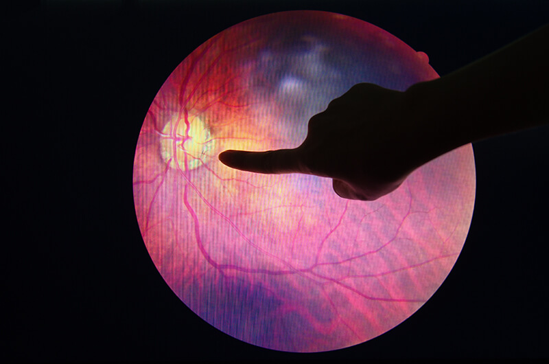
If you have diabetes your body does not use and store sugar properly. High blood-sugar levels can damage blood vessels in the retina, the nerve layer at the back of the eye that senses light and helps to send images to the brain. The damage to retinal vessels is referred to as diabetic retinopathy.
It is important to know that today, with improved methods of diagnosis and treatment, only a small percentage of people who develop retinopathy have serious vision problems. Early detection of diabetic retinopathy is the best protection against loss of vision. You can significantly lower your risk of vision loss by maintaining strict control of your blood sugar and visiting your ophthalmologist regularly.
Diabetic Eye Examinations
When to Schedule an Examination
People with diabetes should schedule examinations at least once a year. More frequent medical eye examinations may be necessary after a diagnosis of diabetic retinopathy. Pregnant women with diabetes should schedule an appointment in the first trimester because retinopathy can progress quickly during pregnancy. If you need to be examined for eyeglasses, it is important that your blood sugar be consistently under control for several days when you see your ophthalmologist. Eyeglasses that work well when the blood sugar is out of control will not work well when the blood sugar is stable.
Rapid changes in blood sugar can cause fluctuating vision in both eyes even if retinopathy is not present.
You should have your eyes checked promptly if you have visual changes that:
- affect only one eye;
- last more than a few days;
- are not associated with a change in blood sugar.
- When you are first diagnosed with diabetes, you should have your eyes checked:
- within five years of the diagnosis if you are 29 years old or younger;
- within a few months of the diagnosis if you are 30 years old and older.
At your Diabetic Eye Exam
To effectively diagnose diabetic eye disease, Orange County Ophthalmology recommends yearly comprehensive diabetic eye examination that includes the following procedures:
- Visual acuity test to check distance and near vision
- A dilated eye examination, which includes the use of an ophthalmoscope. The use of dilating drops allows your doctor to see through the pupil to the retina. Acuity tests alone may not be sufficient to detect diabetic eye disease in its early stages.
- A tonometry test to measure fluid pressure inside the eye.
- A fluorescein angiography test, if more serious retinal changes, such as macular edema, are suspected.
- Optical coherence tomography (OCT) may be used to gain a clearer picture of the retina and its supporting layers.
- Also, an Amsler Grid test can detect early and sometimes subtle visual changes in a variety of macular diseases, including diabetic macular edema.
Diabetic Retinopathy
If you have diabetes your body does not use and store sugar properly. High blood-sugar levels can damage blood vessels in the retina, the nerve layer at the back of the eye that senses light and helps to send images to the brain. The damage to retinal vessels is referred to as diabetic retinopathy.
Types of Diabetic Retinopathy
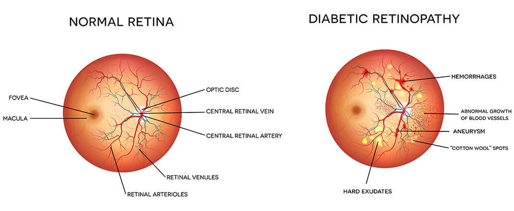
There are two types of diabetic retinopathy: nonproliferative diabetic retinopathy (NPDR) and proliferative diabetic retinopathy (PDR).
NPDR, commonly known as background retinopathy, is an early stage of diabetic retinopathy. In this stage, tiny blood vessels within the retina leak blood or fluid. The leaking fluid causes the retina to swell or to form deposits called exudates.
Many people with diabetes have mild NPDR, which usually does not affect their vision. When vision is affected it is the result of macular edema and/or macular ischemia. Macular edema is swelling or thickening of the macula, a small area in the center of the retina that allows us to see fine details clearly. The swelling is caused by fluid leaking from retinal blood vessels. It is the most common cause of visual loss in diabetes. Vision loss may be mild to severe, but even in the worst cases, peripheral vision continues to function. Macular ischemia occurs when small blood vessels (capillaries) close. Vision blurs because the macula no longer receives sufficient blood supply to work properly.
PDR is present when abnormal new vessels (neovascularization) begin growing on the surface of the retina or optic nerve. The main cause of PDR is widespread closure of retinal blood vessels, preventing adequate blood flow. The retina responds by growing new blood vessels in an attempt to supply blood to the area where the original vessels closed.
Unfortunately, the new, abnormal blood vessels do not resupply the retina with normal blood flow. The new vessels are often accompanied by scar tissue that may cause wrinkling or detachment of the retina. PDR may cause more severe vision loss than NPDR because it can affect both central and peripheral vision.
Proliferative diabetic retinopathy causes visual loss in the following ways:
- Vitreous hemorrhage: The fragile new vessels may bleed into the vitreous, a clear, gel-like substance that fills the center of the eye. If the vitreous hemorrhage is small, a person might see only a few new dark floaters. A very large hemorrhage might block out all vision. It may take days, months, or even years to resorb the blood, depending on the amount of blood present. If the eye does not clear the vitreous blood adequately within a reasonable time, vitrectomy surgery may be recommended. Vitreous hemorrhage alone does not cause permanent vision loss. When the blood clears, vision may return to its former level unless the macula is damaged.
- Traction retinal detachment: When PDR is present, scar tissue associated with neovascularization can shrink, wrinkling and pulling the retina from its normal position. Macular wrinkling can cause visual distortion. More severe vision loss can occur if the macula or large areas of the retina are detached.
- Neovascular glaucoma: Occasionally, extensive retinal vessel closure will cause new, abnormal blood vessels to grow on the iris (colored part of the eye) and block the normal flow of fluid out of the eye. Pressure in the eye builds up, resulting in neovascular glaucoma, a severe eye disease that causes damage to the optic nerve.
How is diabetic retinopathy diagnosed?
A medical eye examination is the only way to detect changes inside your eye. An ophthalmologist [Eye M.D.] can often diagnose and treat serious retinopathy before you are aware of any vision problems. The ophthalmologist dilates your pupil and looks inside of the eye with an ophthalmoscope.
If your ophthalmologist finds diabetic retinopathy, he or she may order color photographs of the retina or a special test called fluorescein angiography to find out if you need treatment. In this test a dye is injected into your arm and photos of your eye are taken to detect where fluid is leaking.
How is diabetic retinopathy treated?
The best treatment is to prevent the development of retinopathy as much as possible. Strict control of your blood sugar will significantly reduce the long-term risk of vision loss from diabetic retinopathy. If high blood pressure and kidney problems are present, they need to be treated.
Laser Surgery: Laser surgery is often recommended for people with macular edema, PDR, and neovascular glaucoma.
For macular edema, the laser is focused on the damaged retina near the macula to decrease the fluid leakage. The main goal of treatment is to prevent further loss of vision. It is uncommon for people who have blurred vision from macular edema to recover normal vision, although some may experience partial improvement. A few people may see the laser spots near the center of their vision following treatment. The spots usually fade with time but may not disappear.
For PDR, the laser is focused on all parts of the retina except the macula. This panretinal photocoagulation treatment causes abnormal new vessels to shrink and often prevents them from growing in the future. It also decreases the chance that vitreous bleeding or retinal distortion will occur.
Multiple laser treatments over time are sometimes necessary. Laser surgery does not cure diabetic retinopathy and does not always prevent further loss of vision.
Vitrectomy: In advanced PDR, the ophthalmologist may recommend a vitrectomy. During this microsurgical procedure, which is performed in the operating room, the blood-filled vitreous is removed and replaced with a clear solution. The ophthalmologist may wait for several months or up to a year to see if the blood clears on its own before performing a vitrectomy.
Vitrectomy often prevents further bleeding by removing the abnormal vessels that caused the bleeding. If the retina is detached, it can be repaired during the vitrectomy surgery. Surgery should usually be done early because macular distortion or traction retinal detachment will cause permanent visual loss. The longer the macula is distorted or out of place, the more serious the vision loss will be.
Laser for Proliferative Diabetic Retinopathy
Diabetic retinopathy is caused by the growth of abnormal new blood vessels in the retina—vessels which leak blood and fluid into the surrounding retina and cause interference with vision. The light from a laser can safely cause these new vessels to regress. In addition to undergoing laser treatments, it is important to maintain the best possible control of your diabetes and to have your retina examined as often as recommended, usually at least once a year. The laser treatments are performed at our office ambulatory surgery center. Usually three sessions are required for each eye.
Arrive one hour before your scheduled treatment in order to have the pupil of your eye dilated with eyedrops. The light of the focusing instrument is bright and somewhat uncomfortable, but not any more so than the instruments during office examinations. A small device similar to an eye cup is placed over your eye. It keeps your eyelids out of the way and holds your eye steady allowing me to focus very precisely on the portions of the retina which need to be treated. This device may feel a little strange, but it doesn’t hurt at all.
Your vision will be blurry for the rest of the day from the eyedrops and also, there will be an afterimage from the bright lights which takes twenty to thirty minutes to fade.
All regular medications can be resumed immediately. Tylenol may be taken for pain, although the laser treatment itself usually does not cause any discomfort. Reading and watching TV are permissible. Vigorous physical activity should be avoided for one day. No special eyedrops are required.